A surgical procedure with the patient’s own bone and tissue is the gold standard, as it does not trigger rejection reactions and integrates best. Surgeons working with own bone and tissue frequently use techniques such as the shell technique according to Khoury and employ own platelet-rich fibrin (PRF) as membranes and clots. Evidence based research fullfilling the Cochrane criteria shows that surgeons working with a patients own bone and tissue have much better results in most of the cases compared to the use of artificial bone and artificial membranes that are offered on the medical market by a multibillion dollar industry.
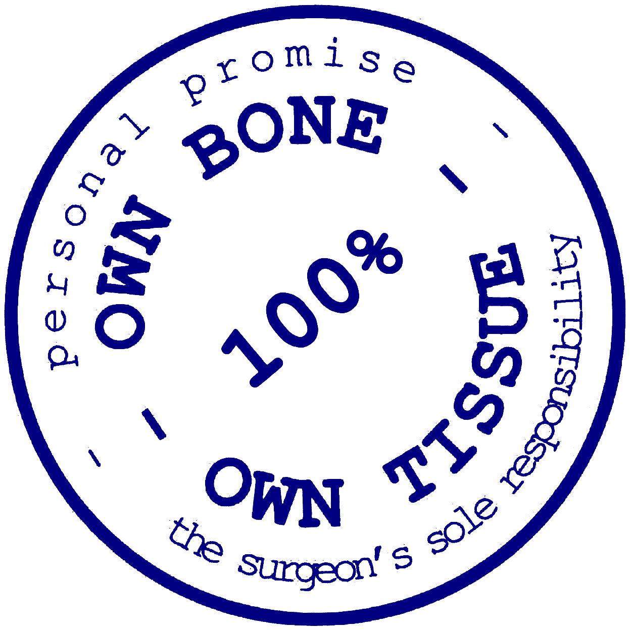
With this seal your surgeon gives you a big promise.
are rigorous, standardized international methods used to conduct systematic reviews of healthcare interventions, ensuring high-quality evidence.
refers in medicine to a diagnostic, therapeutic, or generally scientific procedure that represents the most proven and best solution. New procedures are measured against this gold standard.
Where own bone and own tissue are used:
chin augmentation / genioplasty
complex bone grafting and reconstructive bone surgery
high-end dental implant surgery
simple dental implant surgery
These procedures should be carried out exclusively using own bone and tissue techniques to achieve the most predictable result for each patient.
Key Procedures
Genioplasty
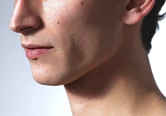
Genioplasty is a precise aesthetic and functional procedure designed to refine the shape, projection and overall balance of the chin. A chin that is small, recessed or asymmetric can disrupt facial proportions and affect the harmony between the lower jaw, lips and nose.
Through controlled advancement, setback, lowering or vertical augmentation, the chin can be reshaped to create a more balanced profile and natural facial symmetry. Surgeons use digital 3D planning and highly accurate surgical techniques to achieve stable contours, functional improvement and predictable, natural-looking results.
Bone Grafting
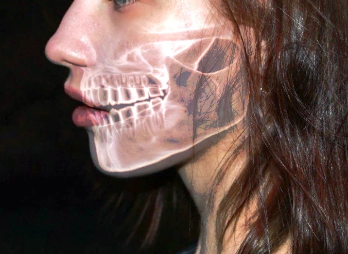
Adequate bone volume is essential for stable implant placement. When bone has been lost due to inflammation, trauma or long-term tooth loss, it can be reconstructed with targeted augmentation techniques.
Depending on the clinical situation, bone grafts, block grafts, internal or external sinus lifts may be used. With precise planning, 3D imaging and minimally invasive approaches, the bone can be safely regenerated to provide a strong foundation for high-quality dental implants. The goal is to achieve functional stability, natural aesthetic reconstruction, and long-term success of the implant-supported restoration.
Implantology
Inflammation, injury, or prolonged tooth loss can cause the jawbone to lose volume, thus compromising the foundation needed for reliable implant treatment. In such cases, however, the bone can be deliberately rebuilt.
A range of proven procedures is available for this purpose: from bone grafts and block bone techniques to internal or external sinus lift methods. Which approach is used depends on the individual clinical findings. Modern DVT imaging and minimally invasive surgical techniques ensure precise, gentle execution throughout the process. The goal is clear: a stable, long-term load-bearing foundation that not only performs functionally but also enables a high-quality aesthetic dental reconstruction for implant treatment that stands the test of time.
Scientific Background and Papers
Fouad Khoury
Fouad Khoury from Germany is a pioneer in advanced bone grafting & sof tissue techniques and has dedicated his career to advancing surgical techniques.
Khoury F. Augmentation of the sinus floor with mandibular bone block and simultaneous implantation: a 6-year clinical investigation. Int J Oral Maxillofac Implants. 1999 Jul-Aug;14(4):557-64. PMID: 10453672.
Khoury F, Hanser T. Mandibular bone block harvesting from the retromolar region: a 10-year prospective clinical study. Int J Oral Maxillofac Implants. 2015 May-Jun;30(3):688-97. doi: 10.11607/jomi.4117. PMID: 26009921.
Khoury F, Hanser T. 3D vertical alveolar crest augmentation in the posterior mandible using the tunnel technique: A 10-year clinical study. Int J Oral Implantol (Berl). 2022 May 13;15(2):111-126. PMID: 35546722.
Khoury F, Hanser T. Three-Dimensional Vertical Alveolar Ridge Augmentation in the Posterior Maxilla: A 10-year Clinical Study. Int J Oral Maxillofac Implants. 2019 Mar/Apr;34(2):471-480. doi: 10.11607/jomi.6869. PMID: 30883623.
Khoury F, Khoury E, Hanser T. 3D Vertical Alveolar Ridge Augmentation in the Partial Edentulous Anterior Maxilla Using the Split-Bone-Block Technique: A 10-year Clinical Study. Int J Periodontics Restorative Dent. 2026 Feb 4;0(0):1-32. doi: 10.11607/prd.7905. Epub ahead of print. PMID: 41637146.
Hanser T, Khoury F. Alveolar Ridge Contouring with Free Connective Tissue Graft at Implant Placement: A 5-Year Consecutive Clinical Study. Int J Periodontics Restorative Dent. 2016 Jul-Aug;36(4):465-73. doi: 10.11607/prd.2730. PMID: 27333003.
Fiorellini J, Lin GH, Rocchietta I, Mojaver S, Aghaloo T, Ahn KM, Al-Nawas B, Araújo M, Shibli JA, Chow J, Faveri M, Fretwurst T, Hürzeler M, Kaigler D, Khoury F, Kim S, Kunrath MF, Lai HC, Messora MR, Sader R, Saleh MHA, Shi J, Jensen SS, Testori T, Urban I, Wu Y, Zad A, Wang HL, Dahlin C. Consensus Report of Group 3 of the 1st Global Consensus for Clinical Guidelines for the Rehabilitation of the Edentulous Maxilla: Advanced Diagnostic Imaging, Augmentation Techniques, and Management of Complications. Clin Oral Implants Res. 2026 Feb;37 Suppl 30(Suppl 30):S68-S80. doi: 10.1111/clr.70079. PMID: 41732077; PMCID: PMC12930124.
Khoury F. The bony lid approach in pre-implant and implant surgery: a prospective study. Eur J Oral Implantol. 2013 Winter;6(4):375-84. PMID: 24570982.
Hanser T, Khoury F. Extraction site management in the esthetic zone using autogenous hard and soft tissue grafts: a 5-year consecutive clinical study. Int J Periodontics Restorative Dent. 2014 May-Jun;34(3):305-12. doi: 10.11607/prd.1749. PMID: 24804281.
Khoury F. Augmentation of the sinus floor with mandibular bone block and simultaneous implantation: a 6-year clinical investigation. Int J Oral Maxillofac Implants. 1999 Jul-Aug;14(4):557-64. PMID: 10453672.
Khoury F, Hanser T. Mandibular bone block harvesting from the retromolar region: a 10-year prospective clinical study. Int J Oral Maxillofac Implants. 2015 May-Jun;30(3):688-97. doi: 10.11607/jomi.4117. PMID: 26009921.
Khoury F, Hanser T. 3D vertical alveolar crest augmentation in the posterior mandible using the tunnel technique: A 10-year clinical study. Int J Oral Implantol (Berl). 2022 May 13;15(2):111-126. PMID: 35546722.
Khoury F, Hanser T. Three-Dimensional Vertical Alveolar Ridge Augmentation in the Posterior Maxilla: A 10-year Clinical Study. Int J Oral Maxillofac Implants. 2019 Mar/Apr;34(2):471-480. doi: 10.11607/jomi.6869. PMID: 30883623.
Khoury F, Khoury E, Hanser T. 3D Vertical Alveolar Ridge Augmentation in the Partial Edentulous Anterior Maxilla Using the Split-Bone-Block Technique: A 10-year Clinical Study. Int J Periodontics Restorative Dent. 2026 Feb 4;0(0):1-32. doi: 10.11607/prd.7905. Epub ahead of print. PMID: 41637146.
Hanser T, Khoury F. Alveolar Ridge Contouring with Free Connective Tissue Graft at Implant Placement: A 5-Year Consecutive Clinical Study. Int J Periodontics Restorative Dent. 2016 Jul-Aug;36(4):465-73. doi: 10.11607/prd.2730. PMID: 27333003.
Fiorellini J, Lin GH, Rocchietta I, Mojaver S, Aghaloo T, Ahn KM, Al-Nawas B, Araújo M, Shibli JA, Chow J, Faveri M, Fretwurst T, Hürzeler M, Kaigler D, Khoury F, Kim S, Kunrath MF, Lai HC, Messora MR, Sader R, Saleh MHA, Shi J, Jensen SS, Testori T, Urban I, Wu Y, Zad A, Wang HL, Dahlin C. Consensus Report of Group 3 of the 1st Global Consensus for Clinical Guidelines for the Rehabilitation of the Edentulous Maxilla: Advanced Diagnostic Imaging, Augmentation Techniques, and Management of Complications. Clin Oral Implants Res. 2026 Feb;37 Suppl 30(Suppl 30):S68-S80. doi: 10.1111/clr.70079. PMID: 41732077; PMCID: PMC12930124.
Khoury F. The bony lid approach in pre-implant and implant surgery: a prospective study. Eur J Oral Implantol. 2013 Winter;6(4):375-84. PMID: 24570982.
Hanser T, Khoury F. Extraction site management in the esthetic zone using autogenous hard and soft tissue grafts: a 5-year consecutive clinical study. Int J Periodontics Restorative Dent. 2014 May-Jun;34(3):305-12. doi: 10.11607/prd.1749. PMID: 24804281.
Sven Nyman and Christoph Dahlin
Sven Nyman and Christoph Dahlin from Sweden laid the biological foundations for guided bone regeneration, which is now used in combination with autologous bone. Dahlin et al. published the fundamental work on bone healing through guided tissue regeneration as early as 1988, and Nyman described the principle of guided tissue regeneration in 1991.
Dahlin C, Linde A, Gottlow J, Nyman S. Healing of bone defects by guided tissue regeneration. Plast Reconstr Surg. 1988 May;81(5):672-6. doi: 10.1097/00006534-198805000-00004. PMID: 3362985.
Dahlin C, Linde A, Gottlow J, Nyman S. Healing of bone defects by guided tissue regeneration. Plast Reconstr Surg. 1988 May;81(5):672-6. doi: 10.1097/00006534-198805000-00004. PMID: 3362985.
Matteo Chiapasco
Matteo Chiapasco from Italy is one of the most prolific researchers in this field. Among other publications, Chiapasco compared autologous onlay bone grafts with alveolar distraction osteogenesis in vertically deficient alveolar ridges, achieving 100% implant survival rates with a follow-up of up to four years. He is considered an expert in complex vertical augmentations.
Chiapasco M, Casentini P, Zaniboni M. Bone augmentation procedures in implant dentistry. Int J Oral Maxillofac Implants. 2009;24 Suppl:237-59. PMID: 19885448.
Chiapasco M, Zaniboni M, Boisco M. Augmentation procedures for the rehabilitation of deficient edentulous ridges with oral implants. Clin Oral Implants Res. 2006 Oct;17 Suppl 2:136-59. doi: 10.1111/j.1600-0501.2006.01357.x. PMID: 16968389.
Chiapasco M, Colletti G, Romeo E, Zaniboni M, Brusati R. Long-term results of mandibular reconstruction with autogenous bone grafts and oral implants after tumor resection. Clin Oral Implants Res. 2008 Oct;19(10):1074-80. doi: 10.1111/j.1600-0501.2008.01542.x. PMID: 18828825.
Chiapasco M, Casentini P, Zaniboni M. Bone augmentation procedures in implant dentistry. Int J Oral Maxillofac Implants. 2009;24 Suppl:237-59. PMID: 19885448.
Chiapasco M, Zaniboni M, Boisco M. Augmentation procedures for the rehabilitation of deficient edentulous ridges with oral implants. Clin Oral Implants Res. 2006 Oct;17 Suppl 2:136-59. doi: 10.1111/j.1600-0501.2006.01357.x. PMID: 16968389.
Chiapasco M, Colletti G, Romeo E, Zaniboni M, Brusati R. Long-term results of mandibular reconstruction with autogenous bone grafts and oral implants after tumor resection. Clin Oral Implants Res. 2008 Oct;19(10):1074-80. doi: 10.1111/j.1600-0501.2008.01542.x. PMID: 18828825.
Carl Misch
Carl Misch from the US was an early pioneer of intraoral bone harvesting. As early as 1995, Misch described the repair of localized severe ridge defects using mandibular bone, and in 1997, he systematically compared various intraoral donor sites prior to implant placement.
Michael Pikos
Michael Pikos from the US established the mandibular block autograft technique in American implantology. In 2005, Pikos published a widely referenced work on mandibular block autografts for alveolar ridge augmentation.
Pikos MA. Atrophic posterior maxilla and mandible: alveolar ridge reconstruction with mandibular block autografts. Alpha Omegan. 2005 Oct;98(3):34-45. PMID: 16273812.
Pikos MA. Mandibular block autografts for alveolar ridge augmentation. Atlas Oral Maxillofac Surg Clin North Am. 2005 Sep;13(2):91-107. doi: 10.1016/j.cxom.2005.05.003. PMID: 16139756.
Pikos MA. Atrophic posterior maxilla and mandible: alveolar ridge reconstruction with mandibular block autografts. Alpha Omegan. 2005 Oct;98(3):34-45. PMID: 16273812.
Miron RJ, Pikos MA. Sinus Augmentation Using Platelet-Rich Fibrin With or Without a Bone Graft: What Is the Consensus? Compend Contin Educ Dent. 2018 Jun;39(6):355-361; quiz 362. PMID: 29847961.
Pikos MA. Atrophic posterior maxilla and mandible: alveolar ridge reconstruction with mandibular block autografts. Alpha Omegan. 2005 Oct;98(3):34-45. PMID: 16273812.
Pikos MA. Mandibular block autografts for alveolar ridge augmentation. Atlas Oral Maxillofac Surg Clin North Am. 2005 Sep;13(2):91-107. doi: 10.1016/j.cxom.2005.05.003. PMID: 16139756.
Pikos MA. Atrophic posterior maxilla and mandible: alveolar ridge reconstruction with mandibular block autografts. Alpha Omegan. 2005 Oct;98(3):34-45. PMID: 16273812.
Miron RJ, Pikos MA. Sinus Augmentation Using Platelet-Rich Fibrin With or Without a Bone Graft: What Is the Consensus? Compend Contin Educ Dent. 2018 Jun;39(6):355-361; quiz 362. PMID: 29847961.
For Surgeons
Become a surgeon with the Seal for Own Bone and Own Tissue
Contact us to use the seal and thereby give your promise. We will add you to the list. You alone will be responsible to keep your promise from then on.
For Patients
Find surgeons with the Seal for Own Bone and Own Tissue
For appointments with surgeons with the Seal for Own Bone and Own Tissue please consult above list.